
XUV imaging
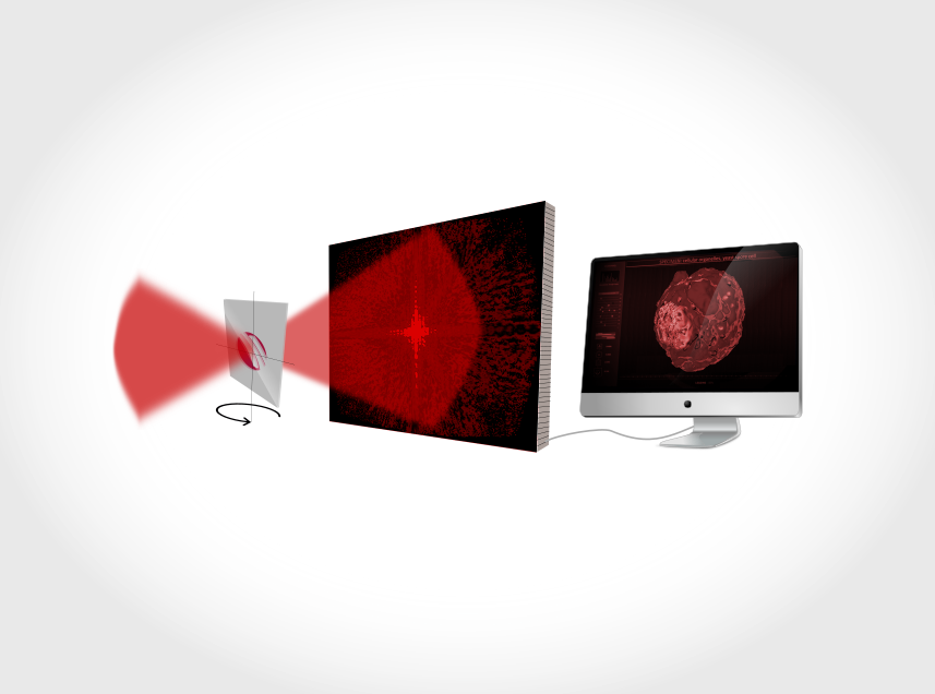
The limits of spatial resolution are classically determined by the wavelength of the employed radiation. Therefore, it is only natural to push forward the utilization of shorter and shorter wavelengths meaning extreme ultraviolet (XUV) and X-ray radiation. Coherent diffractive imaging (CDI) [1], extreme ultraviolet coherence tomography (XCT) [2] and related techniques are variants of X-ray microscopy that have become increasingly important for photon energies where focusing optics are limited in terms of quality and achievable spot size. Samples are illuminated by coherent laser-like beams and the scattered light is detected. The image of the sample is then obtained by means of advanced phase-retrieval algorithms [3]. Ptychography constitutes a generalization of various CDI techniques where the sample is moved with respect to the illuminating beam and multiple diffraction patterns are detected [4, 5]. Employing a table-top XUV source, features as small as 2.5-times the wavelength can be detected [6].
AFS offers fiber-laser-based table-top XUV beamlines with flexible repetition rate providing HHG radiation with photon energies up to 150 eV based on Yb-doped fiber systems. Even higher photon energies up to the soft X-ray regime (e.g. 330 eV [7]) are feasible using Tm-based driving lasers.
View in AR-App

- REFERENCES
- CONTACT
[1] A. Sakdinawat, D. Attwood "Nanoscale X-ray imaging" Nat. Photonics 4, 840–848 (2010).
[2] S. Fuchs et al. "Optical coherence tomography with nanoscale axial resolution using a laser-driven high-harmonic source" Optica 4, 903 (2017).
[3] J. Miao et al. "Beyond crystallography: diffractive imaging using coherent x-ray light", Science 348, 530–5 (2015).
[4] P. Thibault et al. "High-Resolution Scanning X-ray Diffraction Microscopy", Science 321 (2008).
[5] L. Loetgering et al. "Advances in laboratory-scale ptychography using high harmonic sources [Invited]" Optics Express 30 3, 4133-4164 (2022)
[6] G. K. Tadesse et al. "Wavelength-scale ptychographic coherent diffractive imaging using a high-order harmonic source" Sci. Rep. 9, 1–7 (2019).
[7] M. Gebhardt et al. "Bright, high-repetition-rate water window soft X-ray source enabled by nonlinear pulse self-compression in an antiresonant hollow-core fibre" Light Sci. Appl. 1, 36 (2021).
